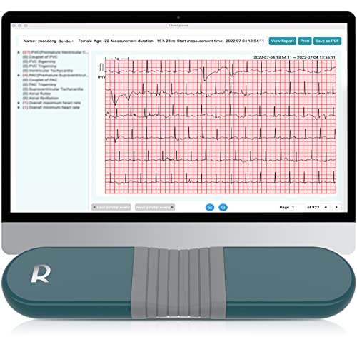
If you are experiencing bouts of tachycardia arrhythmia, they will make you tired all by themselves. Add it to the moderate aortic regurgitation (leakage) and I could see those symptoms occurring. Everyone's response to valve problems is different. Some folks feel symptoms very early on, others don't have much of any symptoms right up to the surgery. You may be sensitive.
Keep your echo results. Particularly compare your Left Venticle (LV) sizes over time (usually expressed as LV diameter measurements). My guess is the LV may be slowly enlarging from stress, but just hasn't reached the upper limits of "normal" by the cardiologist's scale.
If your heart is of small to moderate size, it can grow significantly before it hits those generic measurements on his chart. This early enlargement (hypertrophy) may still cause part of your symptoms. Perhaps the cardiologist is discounting this in your symptoms, because you haven't yet reached the magic number that indicates "enlarged" status
Tachycardia is normal. It just means that your heart increases the speed of its beating when you exercise. This is to provide the extra oxygen needed for your muscles to perform their chores.
Tachycardia arrhythmia (which you have) is pretty much the same thing - except the pulse rate is elevated without the exercise, or continues long after the exercise has ceased. It's a lot of work for your heart. Over time, your heart's rapid beat actually becomes less efficient for transferring oxygen, as the chambers may not fill completely between beats. It causes chest pain and fatigue and may cause lightheadedness, fainting (syncope) or shortness of breath (dyspnea) in some cases.
The tachycardia can even be part of your heart's response to the regurgitation, speeding up the beat to deliver more oxygen short-term. It's not the most common response, but each person's body handles things differently.
Certainly, tachycardia is linked to stress, overwork, and overtiredness. If you're lacking sleep, or have a habit of not knowing when to quit working or playing, you need to correct those things in your lifestyle. Also, cut down on caffeine, which increases your heart rate. Try to lose weight, if you have extra (like most of us do). In general, lowering salt intake is a good idea, especially now that everyone thinks he has to "season" everything as he cooks it, and does so ineptly.
As far as how the leakage works, consider the flow process of the left half of your heart. Relate your heartbeat to its sounds: lub-dub, lub-dub, lub-dub.
lub... Oxygenated blood from the lungs is waiting in the left atrium. The left atrium squeezes, pushing the blood through the mitral valve into the left ventricle. The mitral valve closes, trapping the blood in the left ventricle, and keeping it from leaking back into the left atrium.
...dub. The left ventricle squeezes, and the trapped blood is pushed through the aortic valve, into the aorta. Then the aortic valve closes, so the blood can't flow back from the aorta into the left ventricle.
The percentage of blood from the left ventricle that is pushed out into the aorta is called the ejection fraction (EF), and it's used as a measure of the efficiency of your heartbeat.
The right heart operates similarly, simultaneously with the left heart. The tricuspid valve takes the place of the mitral valve, and the pulmonary valve is in the place of the aortic valve. The right heart is normally less strong and developed, as it only pumps blood to the lungs for oxygen, not to the rest of the body.
The aorta is the large artery leading from the left ventricle of the heart to the rest of the body. Aortic regurgitation (AKA insufficiency or leakage) means that when the aortic valve is supposed to be fully closed in the scenario describe above, blood is still leaking - regurgitating - back through it from the aorta into the LV. This results in there being insufficient blood to completely fill the aorta after the heartbeat. In this way, regurgitation causes, and is often termed, "aortic insufficiency."
In BAV, the leakage is often caused by the valve no longer fitting together tightly to close completely. In some cases, mineral deposits (apatite, which is mostly calcium) interfere with the leaflet movement when the valve tries to close. If those deposits become large enough to block or narrow the path of the bloodflow or thicken the leaflets so that they can't move properly, the flow restriction is called aortic stenosis (AS). Some people with valve disease only develop stenosis. Many people develop both issues.
Aortic insufficiency cuts down on the volume and force of the oxygenated blood being delivered to the brain, heart, and body from the heartbeat as well. Various triggers in the body, including the kidneys, signal that they aren't receiving adequate oxygen. To make up for it, the heart must beat more often and/or more forcefully. Usually, the left ventricle, which is the strongest chamber of the heart, begins to squeeze harder during the beat, and may even "hold" slightly, to keep more of the blood in the aorta after the beat.
From this extra workload, the LV begins to develop muscularly, as any muscle would. This is called ventricular hypertrophy. When it develops evenly, it's termed to be concentric (this is a good thing, if you're going to have LVH at all). Devoted atheletes develop this naturally from their intense training, particularly cyclists.
As the muscle tissue of the LV begins to enlarge, its efficiency does as well. For a while, this is a successful tactic for the heart, and the ejection fraction (EF) returns to normal (50%-65%). This is like you improving your grip by squeezing a tennis ball. If you then try to squeeze juice out of an orange, you will easily get a glassfull from the powerful action of your now-athletic grip.
LVH reaches a plateau from an athlete's exercise, with an ejection fraction that may go up to 75% and still be healthy. Unfortunately, with valve disease, two things tend to occur over time. The valve regurgitation and/or stenosis increases, and the LV growth continues in response. Initially, this will increase your EF, perhaps up to 75% or more, and seem like a good thing.
Unfortunately, as the LV continues to become more bulky and muscular in valve disease, it begins to lose its mechanical advantage because of its size. Consider that your hand has now grown to the size of Arnold Schwartznegger's. Now place a cranberry in your palm, and try to squeeze the juice out of it. You can't get any squeezing force, because the hand is so large in comparison to the fruit. The cranberry just "disappears" in your palm.
When your heart gets to this stage, the EF begins to drop dramatically, and you head toward congestive heart failure. Fortunately, this whole process takes years, and valve surgery is usually done long before this happens.
Although we can't know the exact degree from your post, your aortic regurgitation is rated as moderate, so it's not unlikely that your left ventricle has begun to expand. However, if you have no other heart problems, you could have years to go before the situation is concerning enough to have surgery. A lot of it depends on the speed at which your regurgitation is developing, if at all.
There may be value to more testing, but of the TEE and the cath, I would consider either a cardiac catheterization or a TEE, rather than both. Another option is an MRA (MRI for the heart and arteries). Personally, I'm no fan of stress tests, unless there is a known structural or functional abnormality to be understood. However, your doctor should guide you.
Best wishes,


