Stenosis at 50
Stenosis at 50
It's a lot to adjust to all at once, but you're among a lot of people who've had to come to the same understanding with themselves, and truly understand what you're going through.
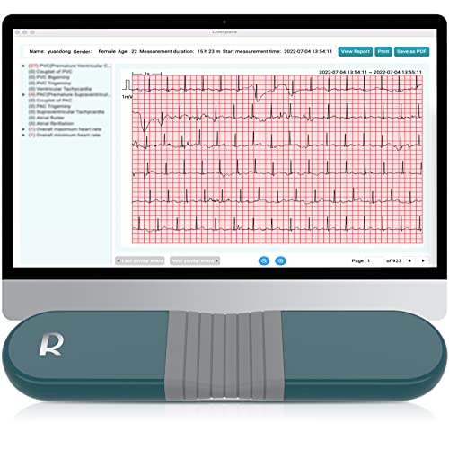
The difference between an electrocardiogram (EKG) and an echocardiogram (ECG) is the same as the difference between a house's electricity and its plumbing. The electrocardiogram measures the electrical impulses from your beating heart. The echocardiogram uses sonar to measure the fluid dynamics going on in your heart as it beats. It provides data to interpret the speed and amount of blood passing through the valves, the sizes of the heart's chambers, and thickness of their walls. It also generates a sonic motion picture of the heart and valves at work, including displaying any leakage or gross anatomical anomalies.
What's likely happening to you (but very slowly), from a nonmedical viewpoint:
When the upper part of your heart squeezes, your mitral valve allows blood to pass from the left atrium into the left ventricle, and keeps it from flowing back. When the lower part your heart squeezes, the aortic valve allows the blood into the aorta (which leads to the brain,the heart muscle, and the rest of the body), and keeps it from flowing back.
The mitral valve has two leaflets that open and close to the passage of blood. Mitral valve prolapse is when the leaflets of the mitral valve are not equally sized, or don't line up perfectly, so the mitral valve doesn't close tightly, and some of the blood going into the ventricle leaks back into the atrium (a.k.a. mitral regurgitation). Mitral valve prolapse is not often serious, and usually doesn't require any surgical action to be taken.
The aortic valve normally has three leaflets. In about 2% of the population, it has only two leaflets, or two of the three are smaller and fused into one. These are termed bicuspid valves. Of those with bicuspid valves, about 1/3 will present with significant problems over their lifetimes.
What usually happens is that a heart that's seemed fine all of one's life will suddenly start generating a murmur. One of the primary times for this to show up is in the early fifties. One theory is that, due to the extra wear they receive, the surfaces of the bicuspid valve leaflets begin to display a chemical signal that the body interprets as damage. The body then attempts to protect the damaged leaflet by coating it with a calcium compound called apatite. As this thickens, it makes the leaflets less flexible, and builds up on the bases of the leaflets, and at their edges to the point where they no longer seal properly.
So several problems are developing. One is that the calcium-coated valve leaflets are no longer flexible, and take more force to open, meaning the ventricle must push the blood harder to get it through. Another is that the buildup of calcium is narrowing the size of the opening that the leaflets allow when they do open. These things in tandem create the symptoms of aortic stenosis, which is usually simply defined as a narrowing of the aortic valve. In response to the body's request for blood, the ventricle now pumps harder and forces the blood through the inflexible, narrow valve opening at a higher pressure and velocity.
The other problem that's developing is that the calcium deposits, building up unevenly at the cusps and the leaflet edges, now cause the valve leaflets to close imperfectly. As well, the increasingly inflexible valve leaflets may not flex enough to fully close any more. These problems allow blood to leak back from the aorta into the ventricle (a.k.a aortic regurgitation). So, as things progress, the ventricle must work even harder to push enough blood into the aorta to fulfill the body's needs, as some of what it pumps out is leaking back in.
All these things work together to cause the left ventricle to enlarge, as any muscle does when it works harder. For a while, that allows it to overcome the problems, but eventually its size interferes with its efficiency. The growth also distorts the valve roots, which in turn increases the amount of regurgitation in both valves.
Where are you in all this? Well, there isn't enough information to hazard a guess from what you've posted. You could very well be years away from any action needing to be taken. However, this is a progressive situation, and you should understand that it will not return to normal without something eventually being done to the valve. There are currently no drugs, nonsurgical therapies, or exercises that can cure or reverse aortic stenosis.
At this point, it would be wise for you to collect your medical records and especially copies of your echocardiogram reports, both for your better understanding later, and for use in determining when you might want to exert pressure on your cardiologist to take action on your behalf. In the Resources Forum, there are also links to places where you can find information about interpreting your own echocardiogram report, to a certain degree.
Fortunately, there are good surgical fixes for the valve. If corrected soon enough, the ventricle will return to its normal size and the heart will return to normal efficiency. There are numerous replacement valves available for this purpose, including several brands of mechanical valves, porcine valves, bovine pericardium valves, and even valves from human cadavers. One of the recurrent themes on this site is discussion of the selection of a valve appropriate to the physical and psychological needs of those who are AVRs-in-waiting.
Take some time to get to know the site and read through some of the information available here. Ask questions as they come up. There are always people here who want to help you.
Be well,


